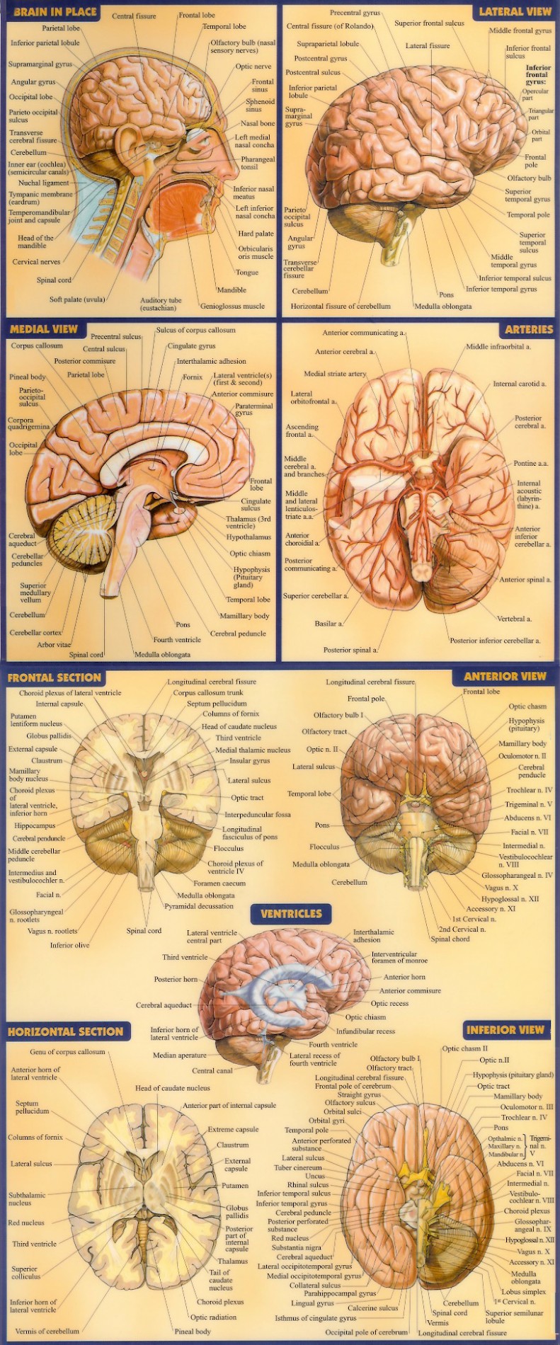
38 human brain diagram with labels
neuron | Definition & Functions | Britannica neuron, also called nerve cell, basic cell of the nervous system in vertebrates and most invertebrates from the level of the cnidarians (e.g., corals, jellyfish) upward. A typical neuron has a cell body containing a nucleus and two or more long fibres. Impulses are carried along one or more of these fibres, called dendrites, to the cell body; in higher nervous systems, only one fibre, the axon ... Parts of Human Eye and Their Functions | MD-Health.com The iris is the area of the eye that contains the pigment which gives the eye its color. This area surrounds the pupil, and uses the dilator pupillae muscles to widen or close the pupil. This allows the eye to take in more or less light depending on how bright it is around you. If it is too bright, the iris will shrink the pupil so that they ...
byjus.com › biology › diagram-of-heartHeart Diagram with Labels and Detailed Explanation - BYJUS The human heart is the most crucial organ of the human body. It pumps blood from the heart to different parts of the body and back to the heart. The most common heart attack symptoms or warning signs are chest pain, breathlessness, nausea, sweating etc. The diagram of heart is beneficial for Class 10 and 12 and is frequently asked in the ...

Human brain diagram with labels
en.wikipedia.org › wiki › Human_eyeHuman eye - Wikipedia The human eye can detect a luminance from 10 −6 cd/m 2, or one millionth (0.000001) of a candela per square meter to 10 8 cd/m 2 or one hundred million (100,000,000) candelas per square meter. At the low end of the range is the absolute threshold of vision for a steady light across a wide field of view, about 10 −6 cd/m 2 (0.000001 candela ... Artificial Neural Network | Brilliant Math & Science Wiki Artificial neural networks (ANNs) are computational models inspired by the human brain. They are comprised of a large number of connected nodes, each of which performs a simple mathematical operation. Each node's output is determined by this operation, as well as a set of parameters that are specific to that node. Structure of the Brain and Their Functions | New Health Advisor Frontal lobe- Associated with planning of speech, reasoning, emotions, problem solving and movement. Occipital lobe- It's associated with visual processing Parietal lobe- It's associated with recognition, movement, orientation, perception of stimuli, speech and memory.
Human brain diagram with labels. byjus.com › biology › human-heartHuman Heart - Anatomy, Functions and Facts about Heart - BYJUS The human heart is one of the most important organs responsible for sustaining life. It is a muscular organ with four chambers. The size of the heart is the size of about a clenched fist. The human heart functions throughout a person’s lifespan and is one of the most robust and hardest working muscles in the human body. human body | Organs, Systems, Structure, Diagram, & Facts human body, the physical substance of the human organism, composed of living cells and extracellular materials and organized into tissues, organs, and systems. Human anatomy and physiology are treated in many different articles. For detailed discussions of specific tissues, organs, and systems, see human blood; cardiovascular system; digestive system, human; endocrine system, human; renal ... bodytomy.com › human-nervous-system-diagramHuman Nervous System Structure and Functions ... - Bodytomy For a detailed study of brain anatomy and its functions, read: diagram of the brain and its functions. Spinal Cord. The spinal cord is a long tubular structure composed of nervous tissue and support cells. It is around 45 cm long in men and 43 cm long in women. It extends from the brain up to the space between the first and the second lumbar ... Brain Structures and Their Functions | MD-Health.com The brain stem consists of midbrain, pons and medulla. Midbrain: The midbrain, also known as the mesencephalon is made up of the tegmentum and tectum. These parts of the brain help regulate body movement, vision and hearing.
Diagram of Human Heart and Blood Circulation in It It beats approximately 72 times per minute, and pumps oxygenated blood to different parts of the body. Atria and Ventricles There are four chambers in your heart that are left atrium, right atrium, left ventricle, and right ventricle. The upper chambers of your heart are atria, whereas the lower chambers are ventricles. Amino acid - Wikipedia Amino acids are organic compounds that contain both amino and carboxylic acid functional groups. Although hundreds of amino acids exist in nature, by far the most prevalent are the alpha-amino acids, which comprise proteins. Only 22 alpha amino acids appear in the genetic code.. Amino acids can be classified according to the locations of the core structural functional groups, as alpha- (α ... Mitochondrion - Wikipedia A mitochondrion (/ ˌ m aɪ t ə ˈ k ɒ n d r i ə n /; pl. mitochondria) is a double-membrane-bound organelle found in most eukaryotic organisms. Mitochondria use aerobic respiration to generate most of the cell's supply of adenosine triphosphate (ATP), which is subsequently used throughout the cell as a source of chemical energy. They were discovered by Albert von Kölliker in 1857 in the ... What Is The Behavioral Perspective? Understanding The ... - BetterHelp It delves into genetics, the immune system, the brain, and the nervous system. This is a perspective that has gotten a lot of attention in the past couple of decades because there are ways of measuring human behavior and explaining it that have to do with tests that can be conducted. It means that often, with this perspective, there is ...
NEUROPHYSIOLOGY Copyright © 2022 Crossing nerve transfer drives sensory ... of the human brain was sufficient to innervate both sides of the body (8). However, how C7 nerve transfer leads to functional recovery of the paralyzed arm in patients remains unknown. In this study, we first established an experimental model in adult mice that replicates nerve transfer-induced functional recovery. Then, we demonstrate Da Vinci Paper Sketches made with Midjourney AI - AI Demos Alien Brain, infographic, paper, drawn by da vinci, blueprint, concept design A pencil sketch of an SR-71 Blackbird drawn in the style of Leonardo da Vinci's flying machine an old page sketch of a staff with an orb of light, leonardo da vinci diagram, highly detailed, sketched on old parchment, desaturated, intricate details -ar 9:16 ... Spinal Cord Cross Section Explained (with Videos) The tracks that move upward are responsible for signals to the brain and the descending tracts send the signals from the brain to neurons throughout the body. 2. Gray Matter In the center of the gray matter you will find the cerebrospinal fluid. The specific horns of the gray matter are responsible for different things. Circulatory System Diagram | New Health Advisor There are different types of circulatory system diagrams; some have labels while others don't. The color blue stands for deoxygenated blood while red stands for blood which is oxygenated. Below you'll see diagram specified to the heart, as well as circulatory system diagram of the whole body: How Does the Human Circulatory System Work? 1. Heart

Drag each label to the correct location on the diagram. label the parts of the brain - Brainly.com
Capillaries: Anatomy, Function, and Significance - Verywell Health The inner layer is made up of endothelial cells with an outer layer of epithelial cells. They are so small that red blood cells need to flow through them single file. It's been estimated that there are 40 billion capillaries in the average human body, and they'd stretch over 100,000 miles if placed in a single line.
Neuroscience - BrainMass Please draw a diagram. Please label and define all lines, arrows, cell types, etc, which are a part of the diagram. ... 7,416 Words; View Details. This book describes the anatomy and function of the human brain. The general focus of the book will be the organization of the brain (i.e., ventricles, the cerebral hemispheres-cerebrum, the ...
Drosophila melanogaster - Wikipedia Drosophila melanogaster is a species of fly (the taxonomic order Diptera) in the family Drosophilidae.The species is often referred to as the fruit fly or lesser fruit fly, or less commonly the "vinegar fly" or "pomace fly". Starting with Charles W. Woodworth's 1901 proposal of the use of this species as a model organism, D. melanogaster continues to be widely used for biological research in ...
› male-human-anatomy-diagramMale Human Anatomy Diagram Pictures, Images and Stock Photos Labeled Anatomy Chart of Male Muscles on White Background Labeled human anatomy diagram of man's full body muscular system from a posterior view on a white background. male human anatomy diagram stock pictures, royalty-free photos & images
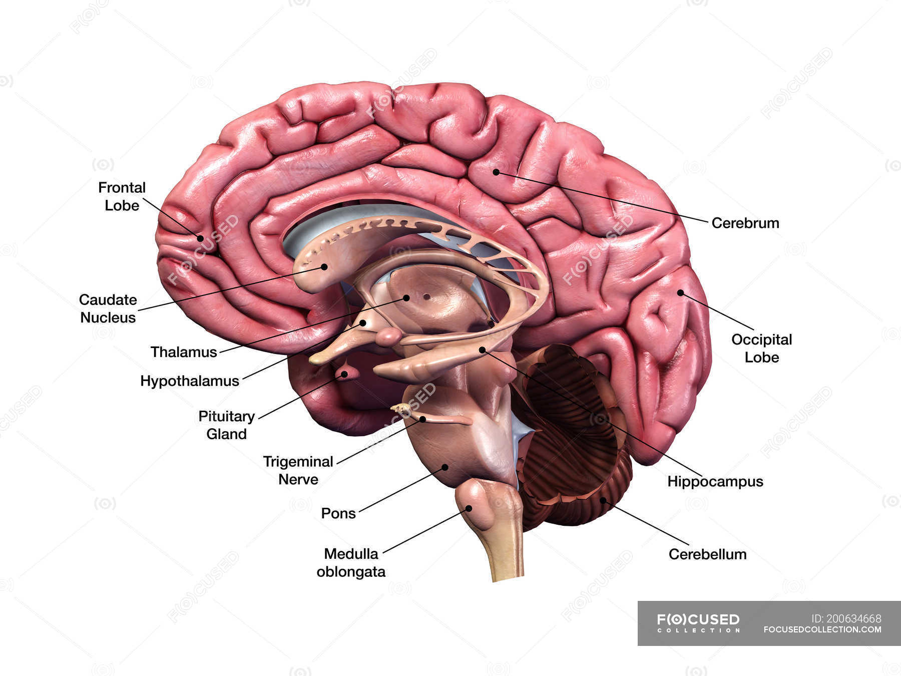
Sagittal section of human brain with labels on white background — trigeminal nerve, physiology ...
Hippocampus: Anatomy, functions and connections | Kenhub The elongated hippocampal structures lie along the longitudinal axis of the brain and form the floor and part of the medial wall of the inferior horns of the lateral ventricles. The hippocampus has many functions, but it is best known for its role in learning and memory . Contents Anatomy Internal structure Dentate gyrus Subicular cortex
› photos › diagram-of-bodyDiagram Of Body Organs Female Pics Pictures, Images ... - iStock Human internal organs Internal organs in woman and man body. Brain, stomach, heart, kidney, medical icon in female and male silhouette. Digestive, respiratory, cardiovascular systems. Anatomy poster vector illustration. diagram of body organs female pics stock illustrations
en.wikipedia.org › wiki › File:Human_skeleton_frontFile:Human skeleton front en.svg - Wikipedia Restructured the image internals by adding layers, changing groupings, and adding meaningful ids and labels so that the image is easier to manipulate programmatically. Also made the labels text elements and gave them ids (it might be possible to generate
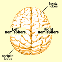
Positions and Functions of the Four Brain Lobes | MD-Health.com The brain is divided into four sections, known as lobes (as shown in the image). The frontal lobe, occipital lobe, parietal lobe, and temporal lobe have different locations and functions that support the responses and actions of the human body. Let's start by identifying where each lobe is positioned in the brain. Position of the Lobes
How Drugs Affect the Brain & Central Nervous System Drugs that can impact GABA levels: benzodiazepines. Norepinephrine: Similar to adrenaline, norepinephrine is often called the "stress hormone," as it speeds up the central nervous system in response to the "fight-or-flight" response. It also homes focus and attention while increasing energy levels.
Structure of the Brain and Their Functions | New Health Advisor Frontal lobe- Associated with planning of speech, reasoning, emotions, problem solving and movement. Occipital lobe- It's associated with visual processing Parietal lobe- It's associated with recognition, movement, orientation, perception of stimuli, speech and memory.

What do the different parts of your brain control... well, here you go! | Human brain anatomy ...
Artificial Neural Network | Brilliant Math & Science Wiki Artificial neural networks (ANNs) are computational models inspired by the human brain. They are comprised of a large number of connected nodes, each of which performs a simple mathematical operation. Each node's output is determined by this operation, as well as a set of parameters that are specific to that node.
en.wikipedia.org › wiki › Human_eyeHuman eye - Wikipedia The human eye can detect a luminance from 10 −6 cd/m 2, or one millionth (0.000001) of a candela per square meter to 10 8 cd/m 2 or one hundred million (100,000,000) candelas per square meter. At the low end of the range is the absolute threshold of vision for a steady light across a wide field of view, about 10 −6 cd/m 2 (0.000001 candela ...





Post a Comment for "38 human brain diagram with labels"