38 parts of the eye without labels
Eye Anatomy: Parts of the Eye and How We See Behind the anterior chamber is the eye's iris (the colored part of the eye) and the dark hole in the middle called the pupil. Muscles in the iris dilate (widen) or constrict (narrow) the pupil to control the amount of light reaching the back of the eye. Directly behind the pupil sits the lens. The lens focuses light toward the back of the eye. Your Eyes (for Kids) - Nemours KidsHealth It is a very important part of the eye, but you can hardly see it because it's made of clear tissue. Like clear glass, the cornea gives your eye a clear window to view the world through. Iris Is The Colorful Part. Behind the cornea are the iris, the pupil, and the anterior chamber. The iris (say: EYE-riss) is the colorful part of the eye. When ...
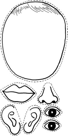
Activity Sheet 1: How the Eyes Work | Human eye diagram, Teaching ... Description Use these simple eye diagrams to help students learn about the human eye. Three differentiated worksheets are included: 1. Write the words using a word bank 2. Cut and paste the words 3. Write the words without a word bank Labels include: eyebrow, eyelid, eyelashes, pupil, iris, and sclera.
Parts of the eye without labels
ear diagram without labels ear diagram without labels Label Parts of the Human Ear we have 9 Images about Label Parts of the Human Ear like Ear Labeling Quiz - Human Anatomy, Label Parts of the Human Ear and also 10 Best Images of Label Ear Diagram Worksheet - Blank Rock Cycle. Here it is: Label Parts Of The Human Ear academic.udayton.edu Anatomy of the Eye | Kellogg Eye Center | Michigan Medicine Structure containing muscle and is located behind the iris, which focuses the lens. Cornea The clear front window of the eye which transmits and focuses (i.e., sharpness or clarity) light into the eye. Corrective laser surgery reshapes the cornea, changing the focus. Fovea The center of the macula which provides the sharp vision. Iris PDF Parts of the Eye Eye Diagram Handout Author: National Eye Health Education Program of the National Eye Institute, National Institutes of Health Subject: Handout illustrating parts of the eye Keywords: parts of the eye, eye diagram, vitreous gel, iris, cornea, pupil, lens, optic nerve, macula, retina Created Date: 12/16/2011 12:39:09 PM
Parts of the eye without labels. Eye Anatomy Detail Picture Image on MedicineNet.com Picture of Eye Anatomy Detail The eye is our organ of sight. The eye has a number of components which include but are not limited to the cornea, iris, pupil, lens, retina, macula, optic nerve, choroid and vitreous. Cornea: clear front window of the eye that transmits and focuses light into the eye. Cornea of the Eye - Definition and Detailed Illustration Cornea Definition. The cornea is the clear front surface of the eye. It lies directly in front of the iris and pupil, and it allows light to enter the eye. Viewed from the front of the eye, the cornea appears slightly wider than it is tall. This is because the sclera (the "white" of the eye) slightly overlaps the top and bottom of the anterior ... Parts of the Eye & Their Function | Robertson Optical and Optometry The different parts of the eye allow the body to take in light and perceive objects around us in the proper color, detail and depth. This allows people to make more informed decisions about their environment. If a portion of the eye becomes damaged, you may not be able to see effectively, or lose your vision all together. The Eyes (Human Anatomy): Diagram, Optic Nerve, Iris, Cornea ... - WebMD The front part (what you see in the mirror) includes: Iris: the colored part. Cornea: a clear dome over the iris. Pupil: the black circular opening in the iris that lets light in. Sclera: the ...
Eye Diagram With Labels and detailed description - BYJUS Iris is the coloured part of the eye and controls the amount of light entering the eye by regulating the size of the pupil. The lens is located just behind the iris. Its function is to focus the light on the retina. The optic nerve transmits electrical signals from the retina to the brain. Pupil is the opening at the centre of the iris. Anatomy of the Eye | Johns Hopkins Medicine Cornea. The clear, dome-shaped surface that covers the front of the eye. Iris. The colored part of the eye. The iris is partly responsible for regulating the amount of light permitted to enter the eye. Lens (also called crystalline lens). The transparent structure inside the eye that focuses light rays onto the retina. Lower eyelid. Labelling the eye — Science Learning Hub Labelling the eye. Use this interactive to label different parts of the human eye. Drag and drop the text labels onto the boxes next to the diagram. Selecting or hovering over a box will highlight each area in the diagram. The human eye has several structures that enable entering light energy to be converted to electrochemical energy. Eye in Cross Section : Anatomy : The Eyes Have It inner layer of posterior wall of eye (see Retina in Cross Section) contains receptors that convert light energy into signals that brain can interpret. Choroid: vascular layer that nourishes outer retina. can be inflamed in autoimmune ("rheumatologic") disorders. Sclera: collagenous outer layer of wall of eye.
Human Eye Ball Anatomy & Physiology Diagram - eMedicineHealth The cornea is located just in front of the iris, which is the colored part of the eye. The main purpose of the cornea is to help focus light as it enters the eye. If one wears contact lenses, the contact lens rests on the cornea. Iris and Pupil. The iris, which is the colored part of the eye, controls the amount of light that enters the eye. PDF Eye Anatomy Handout - National Eye Institute of light entering the eye. Lens: The lens is a clear part of the eye behind the iris that helps to focus light, or an image, on the retina. Macula: The macula is the small, sensitive area of the retina that gives central vision. It is located in the center of the retina. Optic nerve: The optic nerve is the largest sensory nerve of the eye. Learn the Nine Essential Parts of Eyeglasses 1. Rims The rims lend form and character to your eyeglasses—they also provide function by holding the lenses in place. 2. End pieces The end pieces are the small parts on the frame that extend outward and connect the lenses to the temples. 3. Bridge The bridge is the center of the frame that rests on your nose and joins the two rims together. 4. Parts of the Eye - RIT Parts of the Eye Here I will briefly describe various parts of the eye: Sclera The sclera is the white of the eye. "Don't shoot until you see their scleras." Exterior is smooth and white Interior is brown and grooved Extremely durable Flexibility adds strength Continuous with sheath of optic nerve Tendons attached to it The Cornea
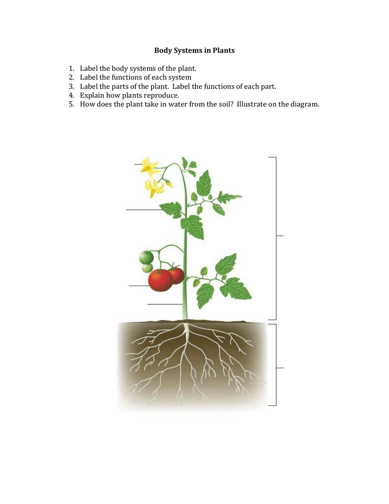
Diagram Of A Flowering Plant With Label - Human Anatomy
Label Parts of the Human Ear - University of Dayton Label Parts of the Human Ear. Select One Auditory Canal Cochlea Cochlear Nerve Eustachian Tube Incus Malleus Oval Window Pinna Round Window Semicircular Canals Stapes Tympanic Membrane Vestibular Nerve. Select One Auditory Canal Cochlea Cochlear Nerve Eustachian Tube Incus Malleus Oval Window Pinna Round Window Semicircular Canals Stapes ...

Horse Pictures to Print | Free printable horse parts diagram with labels | Horses | Pinterest ...
Anatomy of the Eye. Learn about the different parts of the eye. The sclera is a membrane of tendon in the eye, also known as the white of the eye. Rugged and robust, the sclera works to protect the inner, more sensitive parts of the eye like the retina and choroid. It is about 0.03 of an inch thick except for where the four "straight" eye muscles append, where the depth is no more than 0.01 of an inch.
Human Anatomy Review and Question Database
Human eye - Wikipedia The front part is also called the anterior segment of the eye. The eye is not shaped like a perfect sphere, rather it is a fused two-piece unit, composed of an anterior (front) segment and the posterior (back) segment. The anterior segment is made up of the cornea, iris and lens.

Label Parts of the Human Eye - University of Dayton Parts of the Eye Select the correct label for each part of the eye. The image is taken from above the left eye. Click on the Score button to see how you did. Incorrect answers will be marked in red.

Free Unlabeled Eye Diagram, Download Free Unlabeled Eye Diagram png images, Free ClipArts on ...
Quiz: Label The Parts Of The Eye - ProProfs How much did you get to understand about the human eye? Take up this quiz and find out! Questions and Answers. 1. A is pointing to what part of the eye? A. Cornea. B. Optic Nerve.

Blank Ear Diagram | Human ear diagram, Ear anatomy, Ear diagram
Eye anatomy: A closer look at the parts of the eye Eye anatomy: A closer look at the parts of the eye. When surveyed about the five senses — sight, hearing, taste, smell and touch — people consistently report that their eyesight is the mode of perception they value (and fear losing) most. Despite this, many people don't have a good understanding of the anatomy of the eye, how vision works ...
12 Best Images of Eye Parts Worksheet Printable - Electrical Circuit Symbols, Clothes ESL ...
BYJUS BYJUS

Adobe Acrobat Standard Help 7.0 Instruction Manual 7 En
Anatomy of the eye: Quizzes and diagrams - Kenhub Found within two cavities in the skull known as the orbits, the eyes are surrounded by several supporting structures including muscles, vessels, and nerves. There are 7 bones of the orbit, two groups of muscles (intrinsic ocular and extraocular), three layers to the eyeball … and that's just the beginning. There's a lot to learn, but stay calm!
Post a Comment for "38 parts of the eye without labels"