41 chlamydomonas diagram with labels
Animal Cells: Labelled Diagram, Definitions, and Structure Animal Cells Organelles and Functions. A double layer that supports and protects the cell. Allows materials in and out. The control center of the cell. Nucleus contains majority of cell's the DNA. Popularly known as the "Powerhouse". Breaks down food to produce energy in the form of ATP. Gene duplication and evolution in recurring polyploidization ... Feb 21, 2019 · Background The sharp increase of plant genome and transcriptome data provide valuable resources to investigate evolutionary consequences of gene duplication in a range of taxa, and unravel common principles underlying duplicate gene retention. Results We survey 141 sequenced plant genomes to elucidate consequences of gene and genome duplication, processes central to the evolution of ...
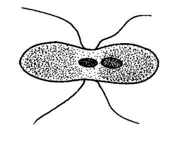
Describe the structure of chlamydomonas with neat labelled diagram ... 1. Chlamydomonas is a simple, unicellular, motile fresh water algae. They are oval, spherical or pyriform in shape. 2. The cell is surrounded by a thin and firm cell wall made of cellulose. 3. The cytoplasm is seen in between the cell membrane and the chloroplast. 4.

Chlamydomonas diagram with labels
Structure of Chlamydomonas (With Diagram) | Genetics In this article we will discuss about the structure of chlamydomonas (explained with labelled diagram). The unicellular green alga Chlamydomonas is haploid with a single nucleus, a chloroplast and several mitochondria (Fig. 9.3). It can reproduce asexually as well as sexually by fusion of gametes of opposite mating types (mt + and mt -). The mating type is controlled by a single nuclear gene. Structure and Diagram of Volvox and Their Functions Thallus structure of volvox is a motile colony with definite shape and number of cells. This habit of thallus is called coenobium. The colony is hollow, spherical or oval in shape and the size of colony is about the size of a pin head. The number of cells in a colony is fixed. Depending upon the species of Volvox the cells can be 500-60,000. Use this labeled diagram of a chlamydomonas cell to A Cancer cells express high levels of a glucose transporter . Code 222. 35. Assume that you have a research hypothesis that using mental imagery (making connections) helps to improve learning among Biology students. You decide to test the prediction that mental imagery made before the test leads to more durable recollection compared to cramming.
Chlamydomonas diagram with labels. Chlamydomonas: Position, Occurrence and Structure (With Diagrams) Chlamydomonas is unicellular, motile green algae. The thallus is represented by a single cell. It is about 20 p,-30|i in length and 20 µ in diameter. The shape of thallus can be oval, spherical, oblong, ellipsoidal or pyriform. The pyriform or pear shaped thalli are common, they have narrow anterior end and a broad posterior end (Fig. 1). LABORATORY 9 - Susquehanna University Look at the video of Chlamydomonas swimming. Then, study the videos of: the mating dance, cells fusing, zygotes swimming. Zygotes are the large cells, and you can just make out that they have 4 flagella as they swim. Figure 1. Labeled diagram of Chlamydomonas. Image from . Figure 2. Life Cycle of Chlamydomonas (With Diagram) - Biology Discussion Each daughter cell develops cell wall, flagella and transforms into zoospore (Fig. 6). The zoospores are liberated from the parent cell or zoosporangium by gelatinization or rupture of the cell wall. The zoospores are identical to the parent cell in structure but smaller in size. The zoospores simply enlarge to become mature Chlamydomonas. Diagram Of Chlamydomonas With Label - Blogger Draw a labelled diagram of chlamydomonas. It is oblong or pyriform in shape. Biological drawings of protista, structure of chlamydomonas,. The anterior end has two tinsel shaped . Shipping a package with ups is easy, as you can print labels for boxes, paste them and even schedule a pickup.
Draw a neat labelled diagram. Chlamydomonas - Shaalaa.com Draw a neat labelled diagram. Chlamydomonas . Maharashtra State Board HSC Science (General) 11th. Textbook Solutions 8018. Important Solutions 19. Question Bank Solutions 5546. Concept Notes & Videos 432. Syllabus. Advertisement Remove all ads. Draw a neat labelled diagram. ... Spirogyra Labelled Diagram Draw a neat diagram of Spirogyra and label the following parts: i. Outermost layer of the cell. ii. Organelle that performs the function of. Each cell of Spirogyra filament is cylindrical and consists of 2 parts: cell wall and protoplast. The cell wall surrounds the protoplast, is protective and consists of. Structure of Chlamydomonas (With Diagram) | Chlorophyta Chlamydomonas is unicellular, motile green algae. The thallus is represented by a single cell. It is about 20 p,-30|i in length and 20 µ in diameter. The shape of thallus can be oval, spherical, oblong, ellipsoidal or pyriform. The pyriform or pear shaped thalli are common, they have narrow anterior end and a broad posterior end (Fig. 1). Schaums Outline Of Genetics [PDF] [1vrels341bno] X chromosomes \ Y chromosome Fig. 1-2. Diagram of diploid cells in Drosophilu melanogaster. CHAP. 11 T H E PHYSICAL BASIS OF HEREDITY 5 CELL DIVISION 1. Mitosis. All somatic cells in a niulticellular organism are descendants of one original cell. the fertilized egg, or zygote. through a divisional process called mitosis (Fig. 1-3).
Lifestyle | Daily Life | News | The Sydney Morning Herald The latest Lifestyle | Daily Life news, tips, opinion and advice from The Sydney Morning Herald covering life and relationships, beauty, fashion, health & wellbeing Chlamydomonas - Wikipedia Chlamydomonas is a genus of green algae consisting of about 150 species all unicellular flagellates, found in stagnant water and on damp soil, in freshwater, seawater, and even in snow as "snow algae". Chlamydomonas is used as a model organism for molecular biology, especially studies of flagellar motility and chloroplast dynamics, biogenesis, and genetics. One of the many striking features of Chlamydomonas is that it contains ion channels that are directly activated by light. Some regulatory sy Chlamydomonas - Meaning, Structure, Life Cycle, Function and FAQs - VEDANTU Given below is the Chlamydomonas structure with labels. The Life Cycle of Chlamydomonas . Chlamydomonas Reproduction is both sexual as well as asexual reproduction. Asexual reproduction takes place by following methods: 1. Zoospore Formation: The protoplast separates from the cell wall as it contracts. The parent cell loses its flagella, or in certain Chlamydomonas species, the flagella are absorbed. Characterization techniques for nanoparticles: comparison and ... The size distribution of their particles depended on the pH of the culture medium. The Ag NP toxicity on the green alga Chlamydomonas acidophila was pH-dependent as shown by the cytotoxicity mediated through the induction of oxidative stress. 227. Pavlopoulou et al. monitored the synthesis of Pt NPs using pH-responsive microgel particles. SAXS ...

Cladocera, water fleas: taxonomy, diversity, anatomy, drawings at GeoChemBio
Biological drawings. Structure of Chlamydomonas. Learning Resources for ... Structure of Chlamydomonas: Next Drawing > Chlamydomonas is the name given to a genus of microscopic, unicellular green plants (algae) which live in fresh water. Typically their single-cell body is approximately spherical, about 0.02 mm across, with a cell wall surrounding the cytoplasm and a central nucleus.
Cells | Special Issue : Chlamydomonas Cell Biology
Solved: Label this diagram of the Chlamydomonas life cycle. | Chegg.com LearnSmart Online for Biology (10th Edition) Edit edition. This problem has been solved: Solutions for Chapter 21 Problem 24TY: Label this. diagram of the. Chlamydomonas life cycle.…. Get solutions. Get solutions Get solutions done loading. Looking for the textbook?

Contoh Explanation Text Photosynthesis - 6 Contoh x
How to make label Diagram of chlamydomonas - YouTube watch: "How to make thumbnail our you tube videos Hindi /urdu haris by #Top2utv" ...
Cells | Special Issue : Chlamydomonas Cell Biology
Diagram of Chlamydomonas angulosa... - Getty Images UNSPECIFIED - CIRCA 2003: Diagram of Chlamydomonas angulosa, Flagellated Protozoan. Drawing. (Photo by DeAgostini/Getty Images)

Biology Champ | Reproduction in organisms
Chloroplast Structure and Function in detail with Labelled Diagram The chloroplasts are the cell organelles which consist of these pigments. The 3 types of pigments present in plants are chlorophyll, carotenoids, and anthocyanins. Chlorophyll imparts the green color to plants. Plastids are membrane-bound cytoplasmic organelles that can be found in the cells of plants and algae.

Chlamydomonas – Parts Labeled | Radesaal the science group
Finance in Germany | Expatica Germany Understanding your money management options as an expat living in Germany can be tricky. From opening a bank account to insuring your family’s home and belongings, it’s important you know which options are right for you.

Chlamydomonas | ClipArt ETC
Solved: Chapter 21 Problem 24TY Solution - Chegg ISBN-13: 9780077388508 ISBN: 007738850X Authors: Sylvia S Mader Rent | Buy. This is an alternate ISBN. View the primary ISBN for: Biology 10th Edition Textbook Solutions.

DRAW IT NEAT : How to draw Plant cell
Morphology of Chlamydomonas (With Diagram) | Algae ADVERTISEMENTS: In this article we will discuss about the external morphology of chlamydomonas. Also learn about its Neuromotor Apparatus, Electron Micrograph, Palmella-Stage with suitable diagram. 1. The organism is an unicellular alga (Fig. 11). 2. The thallus is spherical to oblong in shape but some species are pyriform or ovoid. ADVERTISEMENTS: 3. The cell is […]

Sample Descriptive Lab Report
Microorganisms: Friend and Foe Class 8 Extra Questions ... Oct 11, 2019 · Pull out a gram or bean plant from the field. Observe its roots. You will find round struc¬tures called root nodules on the roots. Draw a diagram of the root and show the root nod¬ules. Answer: Question 2. Collect the labels from the bottles of jams and jellie on the labels. Answer: Do it yourself. Question 3. Visit a dcotor.
Chlamydomonas - JungleKey.fr Image #100
Chlamydomonas | Facts, Structure, Life Cycle, & Classification Chlamydomonas, genus of biflagellated single-celled green algae (family Chlamydomonadaceae) found in soil, ponds, and ditches. Chlamydomonas species can become so abundant as to colour fresh water green, and one species, C. nivalis, contains a red pigment known as hematochrome, which sometimes imparts a red colour to melting snow. The cells of most Chlamydomonas species are more or less oval ...

Antibody producing algae | MMG 233 2013 Genetics & Genomics Wiki | FANDOM powered by Wikia
Eye Diagram With Labels and detailed description - BYJUS Iris is the coloured part of the eye and controls the amount of light entering the eye by regulating the size of the pupil. The lens is located just behind the iris. Its function is to focus the light on the retina. The optic nerve transmits electrical signals from the retina to the brain. Pupil is the opening at the centre of the iris.

draw labelled diagram of chlamydomonas.answer me fast if you want brainliest first comer first ...
Genetic map of the Chlamydomonas reinhardtii plastid genome ... Download scientific diagram | Genetic map of the Chlamydomonas reinhardtii plastid genome. Protein-coding regions are yellow and their exons are labeled with an "E" followed by a number denoting ...

Organisms Formerly Known as Protists at Orange County High School Of The Arts - StudyBlue
Chlamydomonas reinhardtii - an overview | ScienceDirect Topics Chlamydomonas reinhardtii cells are oval shaped, c. 10 μm in length and 3 μm in width, with two flagellae at their anterior end (Figure 1). The cells contain a single chloroplast occupying 40% of the cell volume and several mitochondria. ... Diagram labeling densities in the averaged image. (B) Image average from thin sections of pf14 ...

Chlamydomonas anatomy. It's a unicellular organism, yet it's got a way to make it's energy into ...
Use of corpora in translation studies 1137 Projects 1137 incoming 1137 knowledgeable 1137 meanings 1137 σ 1136 demonstrations 1136 escaped 1136 notification 1136 FAIR 1136 Hmm 1136 CrossRef 1135 arrange 1135 LP 1135 forty 1135 suburban 1135 GW 1135 herein 1135 intriguing 1134 Move 1134 Reynolds 1134 positioned 1134 didnt 1134 int 1133 Chamber 1133 termination 1133 overlapping 1132 newborn 1132 Publishers 1132 jazz 1132 Touch 1132 ...

Comparison of ascorbate peroxidase amino acid sequences. A: Amino acid... | Download Scientific ...
Asymmetric properties of the Chlamydomonas reinhardtii cytoskeleton ... The C. reinhardtii eyespot. (a) A diagram illustrating asymmetric localization of the eyespot relative to the cytoskeleton. Two flagella and four microtubule rootlets extend from a pair of basal bodies at the anterior end of the cell; both the mother basal body (small black oval) and the daughter basal body (small gray oval) are associated with a four-membered rootlet (M4 or D4) and a two ...

LABORATORY 20
Chlamydomonas as a Model Organism - Rice University Chlamydomonas as a Model Organism. Chlamydomonas, a genus of unicellular photosynthetic flagellates, is an important model for studies of such fundamental processes as photosynthesis, motility, responses to stimuli such as light, and cell-cell recognition.C. reinhardi, the most commonly studied species of Chlamydomonas, has a relatively simple genome, which has been sequenced.

DRAW IT NEAT : How to draw Euglena
Labeled Diagram of Spirogyra - QS Study Labeled Diagram of Spirogyra. Plant kingdom. Spirogyra is a sophisticated, filamentous green alga, found in freshwater represented by about 300 species. It is also identified as pond silk, as its fiber burnishes like silk due to the occurrence of mucilage.
Post a Comment for "41 chlamydomonas diagram with labels"