40 labels of a microscope and functions
Microscope With Labeled Parts And Functions Three are 3 or 4 objective lenses on a microscope .These are the some main lenses that are used for specimen visualization. They have a magnification power of 40x-100X. There are about 1-4 objectives lenses attached to one microscope, in which some are front-facing and others are opposite-facing. Lenses have differing magnification Capacities. rsscience.com › stereo-microscopeParts of Stereo Microscope (Dissecting microscope) - Rs' Science Unlike a compound microscope that offers a flat image, stereo microscopes give the viewer a 3-dimensional image that you can see the texture of a larger specimen. [In this image] Examples of Stereo & Dissecting microscopes. Major microscope brands (Zeiss, Olympus, Nikon, Amscope, Omano, Leica …) all produce stereomicroscopes.
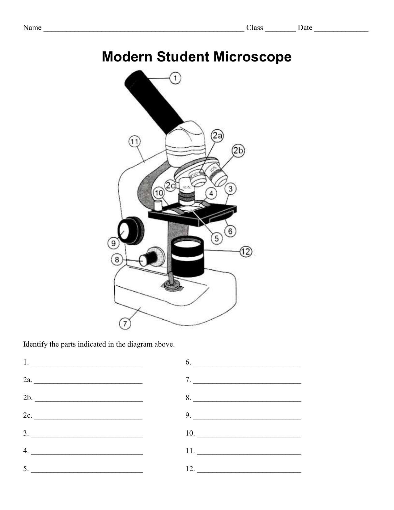
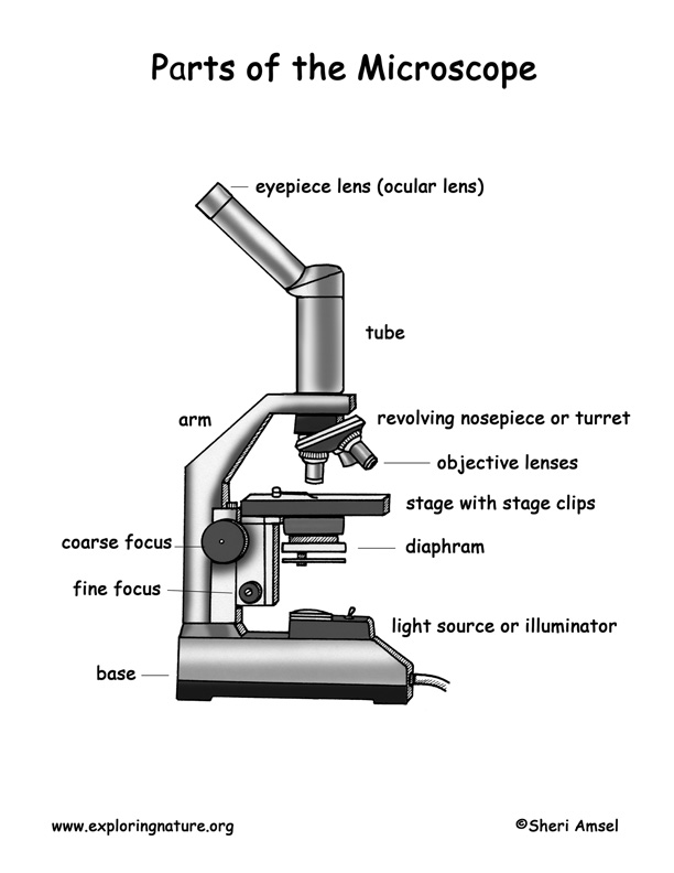
Label Microscope Diagram - EnchantedLearning.com Using the terms listed below, label the microscope diagram. arm - this attaches the eyepiece and body tube to the base. base - this supports the microscope. body tube - the tube that supports the eyepiece. coarse focus adjustment - a knob that makes large adjustments to the focus. diaphragm - an adjustable opening under the stage, allowing ...
Labels of a microscope and functions
Microscope Parts & Functions - AmScope Microscope Parts and Functions Invented by a Dutch spectacle maker in the late 16th century, compound light microscopes use two sets of lenses to magnify images for study and observation. The first set of lenses are the oculars, or eyepieces, that the viewer looks into; the second set of lenses are the objectives, which are closest to the specimen. Microscope Parts, Function, & Labeled Diagram - slidingmotion Microscope parts labeled diagram gives us all the information about its parts and their position in the microscope. Microscope Parts Labeled Diagram The principle of the Microscope gives you an exact reason to use it. It works on the 3 principles. Magnification Resolving Power Numerical Aperture. Parts of Microscope Head Base Arm Eyepiece Lens › 6-label-the-microscopeLabel the microscope — Science Learning Hub Jun 08, 2018 · All microscopes share features in common. In this interactive, you can label the different parts of a microscope. Use this with the Microscope parts activity to help students identify and label the main parts of a microscope and then describe their functions. Drag and drop the text labels onto the microscope diagram. If you want to redo an ...
Labels of a microscope and functions. Microscope Parts and Functions First, the purpose of a microscope is to magnify a small object or to magnify the fine details of a larger object in order to examine minute specimens that cannot be seen by the naked eye. Here are the important compound microscope parts... Eyepiece: The lens the viewer looks through to see the specimen. A Study of the Microscope and its Functions With a Labeled Diagram A Study of the Microscope and its Functions With a Labeled Diagram To better understand the structure and function of a microscope, we need to take a look at the labeled microscope diagrams of the compound and electron microscope. These diagrams clearly explain the functioning of the microscopes along with their respective parts. Microscope parts — Science Learning Hub In this activity, students identify and label the main parts of a microscope and describe their function. By the end of this activity, students should be able to: identify the main parts of a microscope. describe the function of the different parts of a microscope. Download the Word file (see link below) for: background information for teachers. Compound Microscope Parts, Functions, and Labeled Diagram Compound Microscope Definitions for Labels. Eyepiece (ocular lens) with or without Pointer: The part that is looked through at the top of the compound microscope. Eyepieces typically have a magnification between 5x & 30x. Monocular or Binocular Head: Structural support that holds & connects the eyepieces to the objective lenses.
Microscope labeling and functions Flashcards | Quizlet Microscope labeling and functions STUDY Flashcards Learn Write Spell Test PLAY Match Gravity Created by mveet Terms in this set (27) Separates the eyepiece lens from the objective lenses Body Tube Holds the low-power and high-power objective lenses; allows the lenses to rotate for viewing Revolving Nosepiece Magnifies about 4x 22 Parts Of a Microscope With Their Function And Labeled Diagram Invented by a Dutch spectacle maker in the late 16th century, light microscopes use lenses and light to magnify images. Generally a microscope works on the basis of resolution and magnification. The magnifying power of a microscope is an expression of the number of times the object being examined appears to be enlarged and is a dimensionless ratio. › topics › medicine-andConfocal Microscopy - an overview | ScienceDirect Topics John M. Murray, in Methods in Cell Biology, 2013 Abstract. Confocal microscopes are in principle well suited for quantitative imaging. The 3D fluorophore distribution in a specimen is transformed by the microscope optics and detector into the 2D intensity distribution of a digital image by a linear operation, a convolution. Compound Microscope- Definition, Labeled Diagram, Principle, Parts, Uses The optical microscope often referred to as the light microscope, is a type of microscope that uses visible light and a system of lenses to magnify images of small subjects. There are two basic types of optical microscopes: Simple microscopes. Compound microscopes. The term "compound" in compound microscopes refers to the microscope having ...
› doi › 10Expansion microscopy - Science Jan 30, 2015 · Superresolution imaging methods are slower than their diffraction-limited counterparts because they must resolve more voxels per unit volume. ExM achieves this by expanding the voxels physically. ExM achieves the same voxel throughputs as a diffraction-limited microscope, but at the voxel sizes of a superresolution microscope. Microscope- Definition, Parts, Functions, Types, Diagram, Uses Parts of a Microscope 1. Illuminator (Light Source) 2. Diaphragm (Iris) 3. Condenser 4. Condenser Focus Knob 5. Rack Stop 6. Stage 7. Stage Control Knobs 8. Nose Piece 9. Objective Lens 10. Tube (Head) 11. Eyepiece (Ocular Lens) 12. Diopter Adjustment 13. Adjustment Knobs 14. Arm 15. Base 16. Light Switch 17. Brightness Adjustment › internet › portalCanon U.S.A., Inc. | EOS Utility EOS Utility is an application that brings together functions to communicate with the camera. These functions include downloading and displaying images, remote shooting, and camera control for each setting. For download instructions follow the steps below. Have your camera's Serial Number ready before you begin. Compound Microscope Parts - Labeled Diagram and their Functions - Rs ... Basically, compound microscopes generate magnified images through an aligned pair of the objective lens and the ocular lens. In contrast, "simple microscopes" have only one convex lens and function more like glass magnifiers. [In this figure] Two "antique" microscopes played significant roles in the history of biology.
Label And Color The Parts Of Both Microscopes - Ythoreccio
Compound Microscope: Definition, Diagram, Parts, Uses, Working ... - BYJUS A compound microscope is defined as. A microscope with a high resolution and uses two sets of lenses providing a 2-dimensional image of the sample. The term compound refers to the usage of more than one lens in the microscope. Also, the compound microscope is one of the types of optical microscopes. The other type of optical microscope is a ...

Pin on Biología
Parts of a Compound Microscope (And their Functions) List of Microscope Parts and their Functions. 1. Ocular Tubes (Monocular, Binocular & Trinocular) The ocular tubes, are to tubes that lead from the head of the microscope out to your eyes. On the end of the ocular tubes are usually interchangeable eyepieces (commonly 10X and 20X) that increase magnification.
Bacteria - EnchantedLearning.com
Types of Microscopes: Definition, Working Principle, Diagram ... Where, D is the least distinct vision; F is the focal length of the convex lens; Simple Microscope Diagram. Principle of Simple Microscope. The working principle of a simple microscope is that when a sample is placed within the focus of the microscope, a virtual, erect and magnified image is obtained at the least distance of distinct vision from the eye that is held at the lens.
Activity 7 - Brain & Cranial Nerves
Simple Microscope - Parts, Functions, Diagram and Labelling Mirror - A simple microscope has a plano-convex mirror and its primary function is to focus the surrounding light on the object being examined. Lens - The biconvex lens is placed above the stage and its function is to magnify the size of the object being examined.
38 Microscope Diagram To Label - Labels 2021
Compound Microscope Parts, Function, & Diagram - Study.com The base of the compound light microscope is the bottom portion of the compound microscope. It functions to support the entire compound microscope. The base can be set on a table or lab bench, and ...

All Saints Online: Microscope Part Functions
Parts of a microscope with functions and labeled diagram Optical parts of a microscope and their functions The optical parts of the microscope are used to view, magnify, and produce an image from a specimen placed on a slide. These parts include: Eyepiece - also known as the ocular. This is the part used to look through the microscope. Its found at the top of the microscope.

Junior Historians: Awesome Plant & Animal Cells
Labeling the Parts of the Microscope Labeling the Parts of the Microscope This activity has been designed for use in homes and schools. Each microscope layout (both blank and the version with answers) are available as PDF downloads. You can view a more in-depth review of each part of the microscope here. Download the Label the Parts of the Microscope PDF printable version here.
Microscope Labeling Activity
And Parts Quiz Function Of Microscope A stereo microscope has three key parts: Viewing Head/Body that houses the optical components in the upper part of the microscope Microscope Parts and Functions Drag and drop the text labels onto the microscope diagram Palantir Board Of Directors Plant cells are easier to identify because they have a protective structure called a cell wall made ...
Post a Comment for "40 labels of a microscope and functions"